Fig. 2.

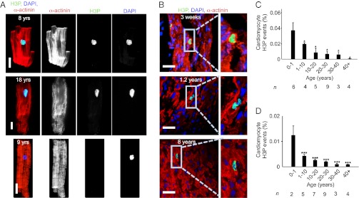
Human hearts show evidence for cardiomyocyte cycling after birth. (A) Isolated cardiomyocytes in M-phase were visualized by immunofluorescent microscopy using antibodies against phosphorylated histone H3 (H3P, green) and α-actinin (red). H3P-negative cardiomyocyte is shown as a control (Lower). (B) H3P-positive cardiomyocytes on tissue sections. (C and D) Quantification of isolated cardiomyocytes by LSC (C) and on tissue sections (D) shows that the percentage of cycling cardiomyocytes decreases with age. Columns represent mean values of the number of hearts investigated per age group (n) that are indicated under each column. One-way ANOVA revealed a significant difference between 0–1 y and the other age groups. *P < 0.05, ***P < 0.001. Scale bar: 25 µm.
