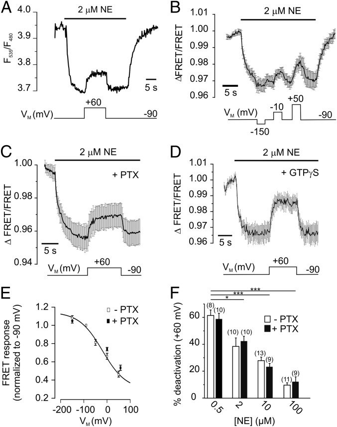
Fig. 1.
The α2AAR is voltage-sensitive. (A) Depolarization reduces the activation of α2AAR-cam. The biosensor was activated with 2 μM NE at −90 mV, reflected as a decrease in the FRET signal. Subsequent depolarization to +60 mV deactivates α2AAR-cam, indicated by a rise in the FRET ratio. The FRET ratio was calculated as F535/F480 (see Fig. S1 for detailed properties of F485 and F535). (B) The FRET response of α2AAR-cam to NE was measured at various membrane potentials as indicated. The trace represents mean ± SEM, n = 4. (C) Voltage-dependence of α2AAR-cam after uncoupling of G proteins by PTX (30 ng/mL for 3 to 5 h, n = 11) or irreversible activation of G proteins by GTPγS (500 μM, n = 6) (D). (E) Normalized FRET amplitudes from α2AAR-cam (FRET/FRET−90mV) were plotted against the membrane potential VM and fitted to a Boltzmann function giving rise to a V0.5 = −20 mV (n = 5–10 cells per datapoint; control: filled squares; PTX: open squares). (F) The magnitude of receptor deactivation at +60 mV was attenuated at high NE concentrations in both control and PTX-treated cells. Data are presented as mean ± SEM of n individual cells (*P = 0.0101, ***P < 0.001).

