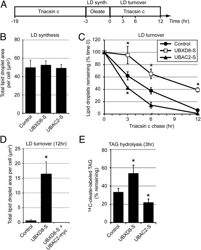
Fig. 3.
LD-localized UBXD8 inhibits lipolytic degradation of LDs. (A) Timeline for oleate (200 µM) and triacsin C treatments (10 µM) for the experiments shown in B–E. (B–D) LD area per cell was quantified at time 0 to measure LD synthesis (B) or at the indicated time points to measure LD turnover (C and D) in transfected HeLa cells stained with BODIPY 493/503. (E) HeLa cells expressing the indicated constructs were treated according to the timeline shown in A, except that 14C-oleate (0.3 µCi) was included during the oleate pulse. Cellular lipids from the 0 and 3 h time points were extracted and separated by TLC, and the amount of 14C-oleate–labeled TAG was determined by scintillation counting. All graphical data are quantified as mean ± SEM. An asterisk indicates a significant difference (P < 0.05, t test) from the control based on three independent biological replicates. (Scale bars: 10 μm.)

