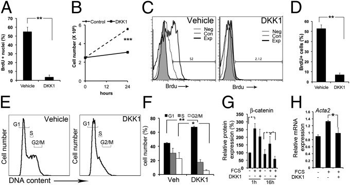
Fig. 2.
DKK-1 binds to myofibroblasts and blocks proliferation by G1/S cell-cycle arrest in vitro. (A) Graph showing BrdU nuclear incorporation in quiescent myofibroblasts stimulated for 3 h with 3% FCS and DKK-1 or vehicle. (B) Coulter-counted kidney quiescent myofibroblasts stimulated for 24 h with 3% FCS and DKK-1 or vehicle. (C and D) Flow cytometric plots (C) and graph (D) showing BrdU uptake in myofibroblasts stimulated for 3 h with 3% FCS and DKK-1 or vehicle. (E and F) Propidium iodide DNA content plots (E) and graph (F) showing quiescent myofibroblasts stimulated for 24 h with 3% FCS and DKK-1 or vehicle. (G) The effect of DKK-1 on cytoplasmic and nuclear β-catenin protein. Serum increases β-catenin, an effect not modulated by DKK-1 at 1 h, but DKK-1 markedly reduces β-catenin levels at later time points. (H) qPCR data showing myofibroblast expression of Acta2 after treatment with FCS or FCS + DKK-1. *P < 0.05, **P < 0.01, ***P < 0.001. n = 4 per group. Error bars indicate SEM.

