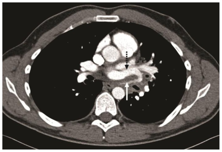
Figure 3.
32-year-old male with complex congenital pulmonary vein varix. ECG gated MDCT performed on 64 slice CT scanner: Contrast enhanced axial image at the level of pulmonary artery origin obtained during pulmonary arterial phase using a mediastinal vascular window shows tortuous vessel in the posterior mediastinum passing between left superior pulmonary vein and descending aorta (solid white arrow) and draining into the left atrium via the left superior pulmonary vein (dotted black arrow).
Protocol: 40mAs, 120kV, 0.7mm slice thickness, 80ml Ultravist 370

