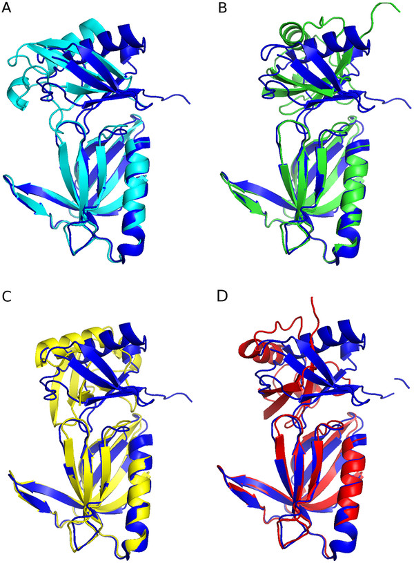
Figure 2.
Best structures of the docked Rpn13-ubiquitin complex structures compared with the high resolution structure. The best structures, concerning their RMSD at the interface compared with [PDB:2Z59] of all methods are overlayed onto [PDB:2Z59] (dark blue). Structures based on limited proteolysis are colored in light blue, based on chemical shift perturbations in green (CSP-50) and yellow (CSP-0), and based on CPORT predictions are colored in red. (A) Comparison of LP/MS and with [PDB:2Z59] (2.85 Å to target). (B) Comparison of CSP-50 with [PDB:2Z59] (6.70 Å to target). (C) Comparison of CSP-0 with [PDB:2Z59] (10.13 Å to target). (D) Comparison of CPORT with [PDB:2Z59] (7.99 Å to target). This figure was generated with PyMOL [32].

