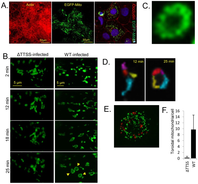
Figure 2. EPEC induces toroidal-shaped mitochondria in polarised TC-7 cells.
Expression of a mitochondrial-targeted EGFP protein in polarised TC-7 cells shows a high level of transfection efficiency with occludin staining pattern confirming polarisation (A). Live cell imaging of EGFP-labelled mitochondria was performed in polarised TC-7 cells infected with a type three secretion system (ΔTTSS) defective mutant (espA) or wild type (WT) EPEC (B). Selected image captures are given with the infection time (left) (B). Captured images show the spatial organisation of mitochondria in cells infected with the two mutants. Arrows show the doughnut shaped (toroidal) mitochondria (B). Toroidal mitochondria were derived from a fusion of individual mitochondria (pseudocoloured; D). Late stage infected cells (60 min) containing many toroidal mitochondria (pseduocoloured red; E), quantified in (F). Toroidal-shaped mitochondria were defined as a continuous ring surrounding a central void. Data represents the mean ± SD, n = 3.

