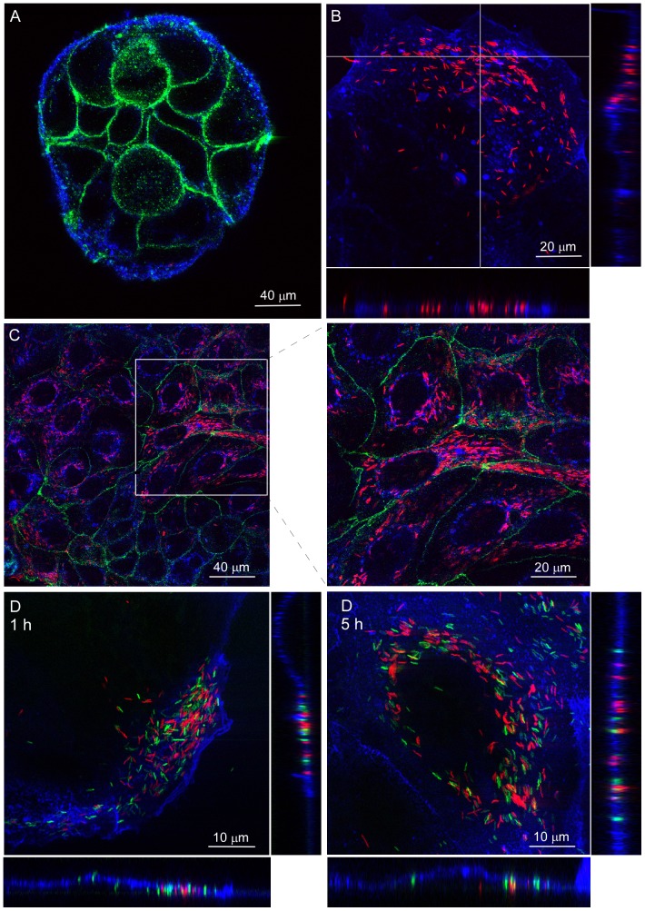
Figure 1. C. jejuni invades polarized Caco-2 islands via subvasion with high efficiency.
Confocal laser microscopy on non-infected and C. jejuni-infected islands of polarized Caco-2 cells. (A) Uninfected island of Caco-2 cells stained with the membrane marker WGA-Alexa fluor633 (Blue) and an anti-occludin antibody (Green) showing the presence of tight junctions. (B) Caco-2 cells (Blue) at 1 h of infection in DMEM showing C. jejuni strain 108p4 (Red) mostly located at the basal side of cells near the edge of the island of polarized cells. (C) Caco-2 cells (Blue) at 5 h of infection in DMEM demonstrating intracellular C. jejuni strain 108p4 (Red) at the center of the island of cells with tight junctions (Green). (D). Polarized Caco-2 cells (Blue) infected (1 h and 5 h) with a mixture of C. jejuni strains 108p4 (Red) and 81–176 (Green) showing invasion of Caco-2 cells by both strains. Transversal optical sections of the cells are depicted at the bottom of each panel to show the location of the bacteria relative to the cell basis.

