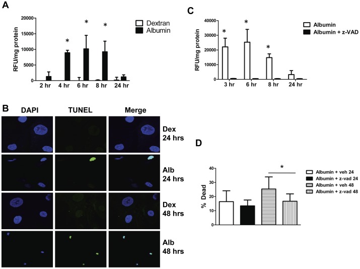
Figure 3. Albumin exposure upregulates pro-apoptotic pathways in podocytes.
A, Activated caspase 3/7 activity normalized to total cellular protein in podocytes treated with 5 mg/ml albumin (closed bars) or 5 mg/ml dextran (open bars) as an oncotic control for the indicated amounts of time. * denotes P<0.0001 compared to corresponding dextran treated control. B, TUNEL staining of cultured podocytes treated with 5 mg/ml dextran (Dex) or 5 mg/ml albumin (Alb). Nuclei are stained blue with DAPI. TUNEL-positive nuclei are green. C, Caspase 3/7 activity normalized to total cellular protein in podocytes treated with 5 mg/ml albumin or 5 mg/ml albumin +50 µM z-VAD, a pan-caspase inhibitor. z-VAD largely abrogated caspase activity (*, P = 0.01 versus albumin+z-VAD). D, Percentage of dead cells measured using the trypan blue assay in podocytes treated with 5 mg/ml albumin alone (open bar) or 5 mg/ml albumin +50 µM z-VAD (closed bar) for 24 hrs or 5 mg/ml albumin alone (horizontal hatched bar) or 5 mg/ml albumin +50 µM z-VAD for 48 hrs (vertical hatched bar). * denotes P = 0.001 compared to albumin+z-VAD at 48 hrs.

