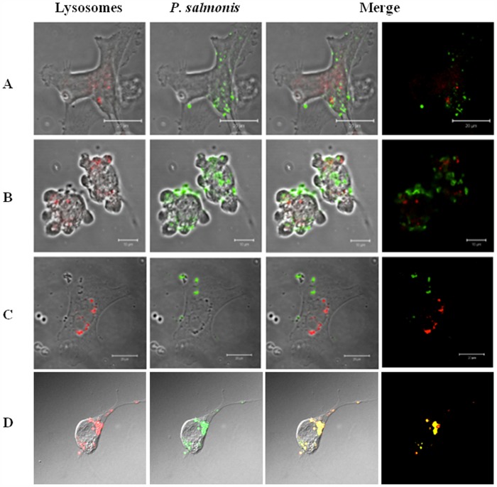
Figure 4. Confocal Laser Scanning Microscopy of P. salmonis infection on three cell lines showing the escape of phagosome-lysosome fusion.
The immunofluorescence was made 5 days post-infection. Lysosomes were stained in red with LysoTracker Red reagent and P. salmonis was detected with a FITC conjugated antibody. A: CHSE-214 cell line infected with P. salmonis. B: Sf21 cell line infected with P. salmonis. C: RTS11 cell line infected with P. salmonis. D: CHSE-214 cell line infected with formaldehyde-inactivated P. salmonis, the immunofluorescence stain was made at 48 hours after infection.

