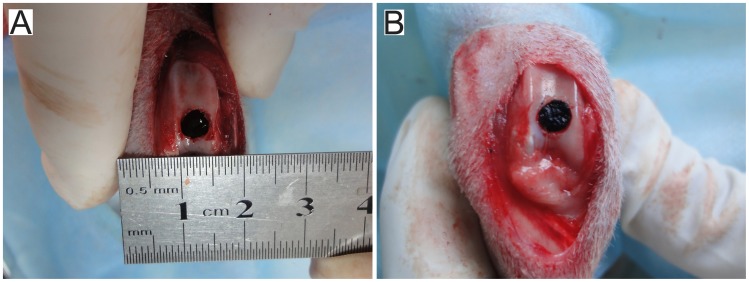
Figure 2. Implantation surgery.
(A) An osteochondral defect (diameter = 5 mm; depth = 6 mm) generated in the rabbit knee patellofemoral groove surface. (B) Prepared autogeneic tissue-engineered osteochondral composite inserted into the defect, and the surface of the composite flushed with the articular surface.

