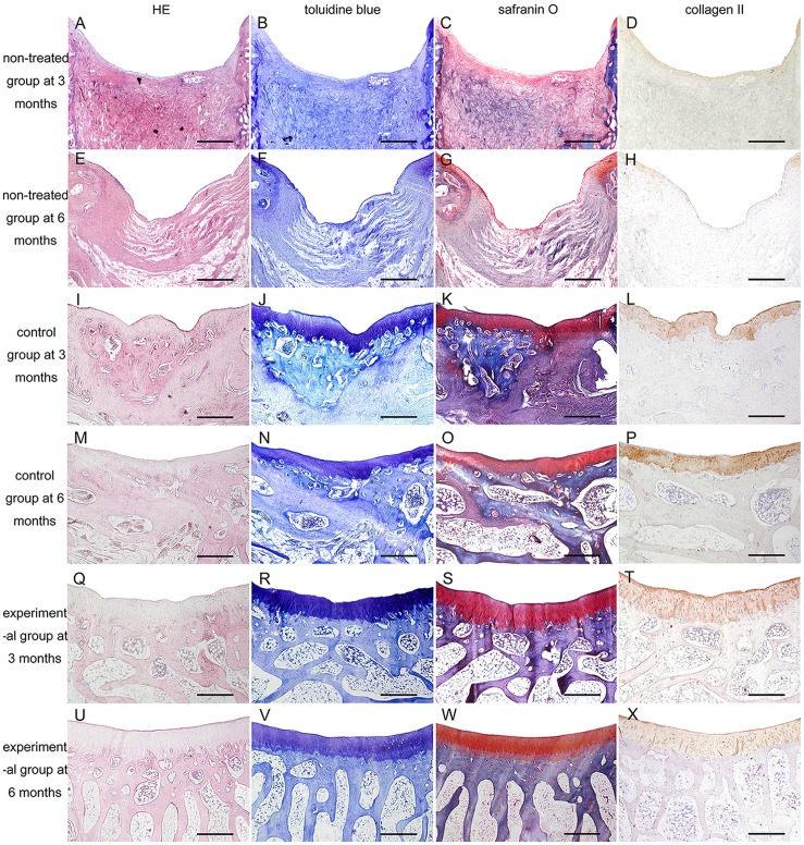
Figure 10. Histological and immunohistochemical analyses of repaired osteochondral tissue.
(A,E,I,M,Q,U) H&E staining. (B,F,J,N,R,V) Toluidine blue staining. (C,G,K,O,S,W) Safranin O staining. (D,H,L,P,T,X) Immunohistochemical staining for collagen type II. (A,B,C,D) Non-treated group defects at 3 months after surgery. (E,F,G,H) Non-treated group defects at 6 months. (I,J,K,L) Control group defects at 3 months. (M,N,O,P) Control group defects at 6 months. (Q,R,S,T) Experimental group defects at 3 months. (U,V,W,X) Experimental group defects at 6 months. Scale bars = 1 mm.

