Figure 1.
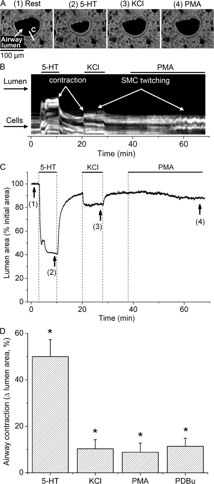
Contractile response of airway SMCs to 5-HT, KCl, and phorbol esters. (A) Representative phase-contrast images showing an airway before stimulation (1) and after stimulation with 0.5 µM 5-HT (2), 50 mM of isosmotic KCl sHBSS (3), and 10 µM PMA (4), taken at the times indicated by arrows and corresponding numbers in the trace shown in C. (B) Line scan obtained from phase-contrast images at the region indicated by a line on image 1 in A, showing the movement of a few cells in the periphery of the airway wall in response to stimulation. 5-HT induced a strong and sustained movement of cells (light gray lines) toward the airway lumen (black), whereas KCl and PMA induced transient cell movements (SMC twitching, observed as striated white lines) and small sustained cell movements. (C) Trace showing the changes in total airway lumen cross-sectional area during superfusion with 5-HT, KCl, and PMA (upper lines). Washout of stimuli was performed by superfusion of lung slices with sHBSS. Sustained airway contraction induced by PMA was small and developed slowly as compared with airway contractions induced by 5-HT and KCl. (D) Summary of sustained airway contraction measured as the decrease in lumen area at 8 min after the addition of 5-HT or KCl, or at 30 min after the addition of PMA or 5 µM PDBu. Data are means ± SEM of seven airways (each from a different lung slice) from three mice. *, different from baseline airway lumen area (P < 0.01). A time-lapse movie showing the airway responses to 5-HT, KCl, and PMA from the representative experiment presented here is shown in Video 1.

