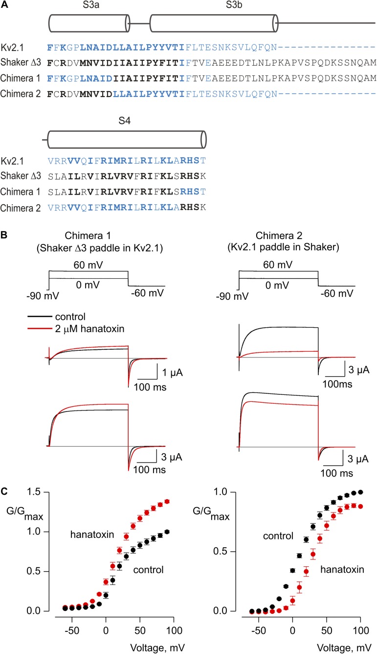
Figure 7.
Influence of hanatoxin on paddle chimeras between Kv2.1 and Shaker Kv channels. (A) Sequence alignments for the S3-S4 regions of Kv2.1 (blue), Shaker Δ3 (black), and two chimeras wherein the S3b-S4 paddle motifs were swapped between the two Kv channels. Conserved residues are shown using bold lettering. (B) Voltage-activated Kv channel currents for two chimeras in the absence (black) and presence (red) of hanatoxin. The top sets of traces are for depolarization to 0 mV, whereas the lower are for depolarization to +60 mV. The gray line indicates the level of zero current. (C) Normalized G-V relations in the absence (black circles) and presence of (red circles) 2 µM hanatoxin. The external solution contained 50 mM K+. Error bars indicate SEM (n = 5).

