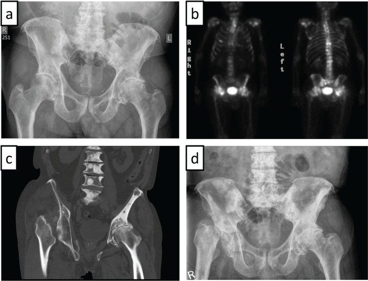
FIGURE 1.
(a) Plain radiography of the pelvis shows osteoarthritic changes of both hips, with joint-space narrowing particularly on the left side. (b) Technetium bone scan reveals multiple foci of increased activity compatible with bone metastases. Notably, the configuration of the right hip is unusual. (c) Near-complete resorption of the right femoral head, with superior displacement of the right femur as seen on computed tomography imaging. (d) Plain radiography of the pelvis reveals fragmentation of the right femoral head, with subluxation of the right femur, multifocal sclerotic metastases throughout the pelvis and lumbar spine, and severe superior migration osteoarthritis of the left hip.

