
Fig 2.
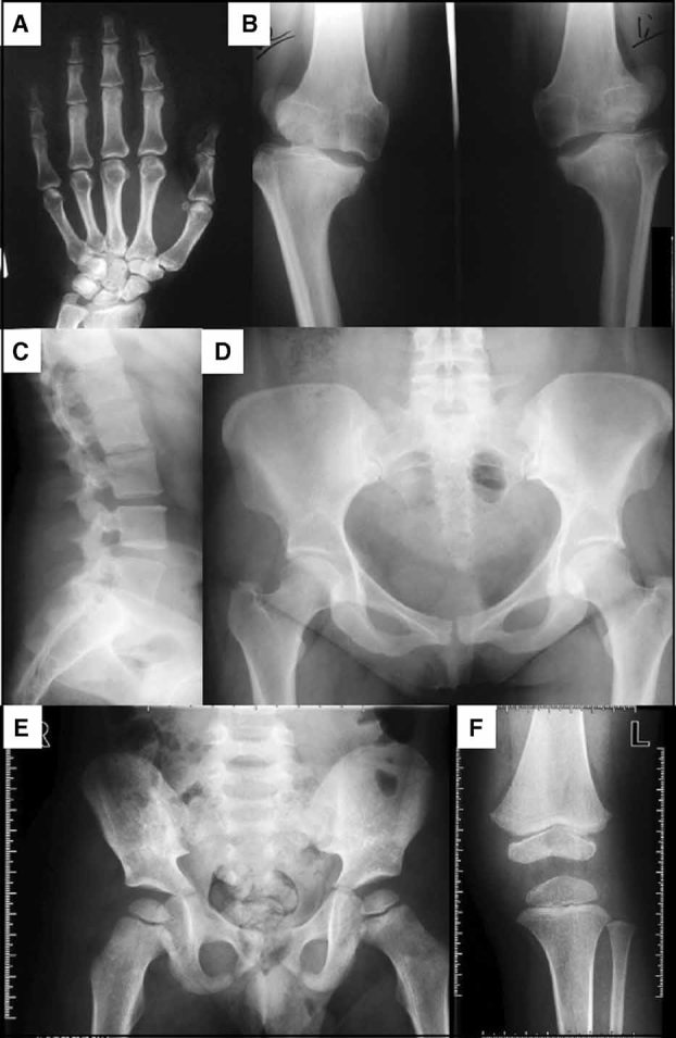
Radiographs of the proband (individual III-4, panels A–D) in Family 1 at 33 years of age and her eldest son (individual IV-6, panels E,F) at 3 years of age. The mother's radiographs showed (A) normal hand, (B) knees with bilateral genu varus, irregular joint surfaces, and decreased joint space, especially of the right knee, (C) lateral spine with irregular endplates of the vertebral bodies in the thoracolumbal region and (D) relatively normal hips. Radiographs of her 3-year-old son showing (E) hips and (F) the left knee. The radiographs were consistent with a diagnosis of mild MED, characterized by delayed ossification of the epiphyses. The hips were relatively spared but had small proximal femoral epiphyses, while the knees had small femoral and tibial epiphyses.