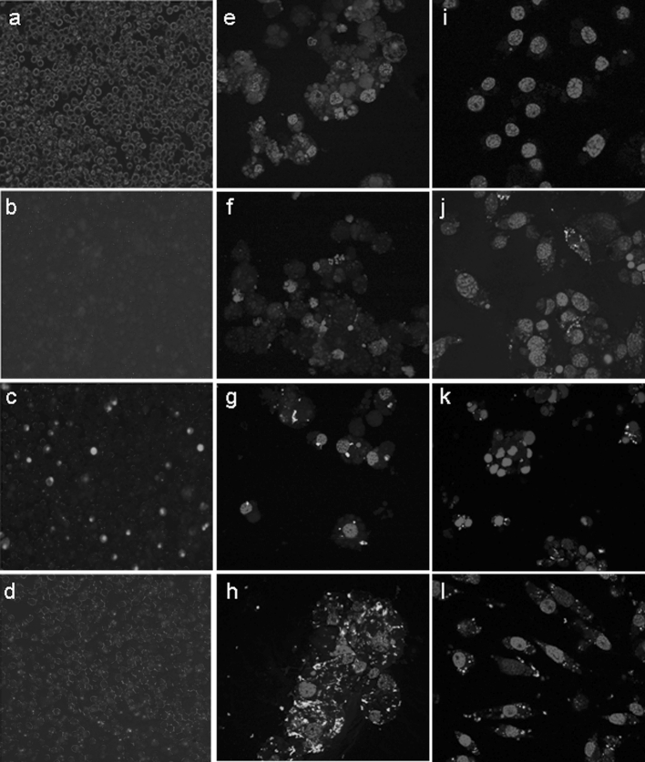
Fig. 1.
Images of tick cells 24 h after addition of fluorescent siRNA (a–h) or dsRNA (i–l) in the presence or absence of a transfection reagent. a–d photomicrographs of BDE/CTVM16 cells taken at ×100 magnification with simultaneous normal transmitted light and UV reflected light to facilitate counting of fluorescent (green siRNA) and non-fluorescent cells. e–h confocal images of IRE/CTVM19 cells taken at ×630 magnification; cell nuclei are stained blue, while the siRNAs are green. i–l confocal images of IDE8 cells taken at ×630 magnification; cell nuclei are stained blue, while the long dsRNAs are green. a, e, i show untreated control cells; b, f, j show cells to which siRNA or dsRNA alone was added; c, g, k show cells to which siRNA or long dsRNA mixed with Lipofectamine 2000 was added; d, h, l show cells to which siRNA or long dsRNA mixed with Xtreme was added. (Color figure online)

