
FIG. 10.
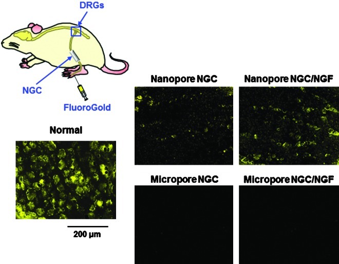
Fluorescent micrographs following FluoroGold (FG) retrograde tracing in the DRGs of the NGC-implanted group 1 week after FG injection (×100). The Nanopore NGC/NGF group contained more FG-labeled neuron cells compared with the Nanopore NGC group, indicating that a greater number of nerve fibers connecting the defect stumps were present. However, FG-labeled cells in the DRGs were not detected in the Micropore NGC groups, even for the NGF-immobilized NGC group. DRG, dorsal root ganglions. Color images available online at www.liebertpub.com/tec