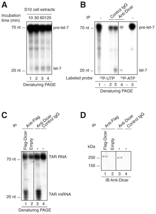
Figure 1. Detection of human Dicer activity in HEK293 cells.
(A) S10 protein extracts were incubated in the presence of a 32P-UTP labeled human let-7a-3 pre-miRNA substrate for the indicated period of time. (B) Endogenous Dicer immune complexes were incubated with a 32P-UTP or 32P-ATP labeled human let-7a-3 pre-miRNA substrate for 60 min. Lanes 1 and 5 represent the untreated probe (−). (C) Anti-Flag immune complexes derived from cells overexpressing Flag-Dicer, or transfected with empty plasmid, and endogenous Dicer immune complexes derived from untransfected HEK293 cells (−) were incubated in the presence of 32P-UTP-labeled TAR RNA substrate for 60 min. (A–C) The reactions were analyzed by denaturing PAGE and autoradiography. A 10-nt RNA ladder was used as a size marker. (D) The immune complexes from (C) were analyzed for the presence of Dicer protein by 7% SDS-PAGE and immunoblotting using anti-Dicer antibody (9).

