
FIGURE 4.2.
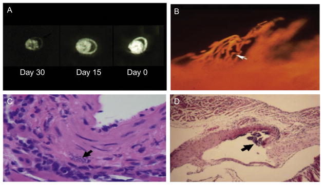
(A) Coronary perfusion of mouse hearts as determined by autoradiographic imaging utilizing the fatty acid analog 19-iodo-3,3,-dimethyl-18 nonadecenoic acid (DMIVM). A: uninfected normal mouse with normal perfusion. B: perfusion in a mouse infected for 15 days. Note the reduced perfusion C: perfusion in a mouse infected for 30 days demonstrating a marked reduction in perfusion (taken from Tanowitz, 1992). (B) Microfil injection of the coronary vasculature of an A/J mice 15 days post-infection with the Tulahuen strain of T. cruzi demonstrating a section through the subendocardium of the atrium showing saccular microaneurysms and vasospasm (Rossi et al., 2010). (C) Pseudocyst in the wall of a blood vessel (Tanowitz et al., 2009). (D) Vasculitis of a large blood vessel obtained from an infected mouse (Tanowitz et al., 2009).