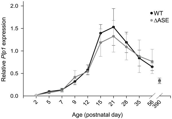
Fig. 4.
qRT-PCR analysis of Plp1 gene expression in WT and ΔASE male mice from P2 to P390. A custom designed primer/probe set for Plp1 was used, which detects all splice variants (Plp and Dm20). Results were obtained using the 2−ΔΔCT method, with β-actin as the reference gene. Results are plotted as the mean level ± SD of Plp1 (Plp and Dm20 mRNA combined) relative to that from a uniform control (pooled brain mRNA from P21 WT mice) for each timepoint/genotype (n ≥ 4 for P2-P7; n ≥ 3 for P9-P390). While there tended to be a slight reduction in the level of Plp1 gene expression in ASE-deleted (ΔASE) mice compared to WT littermates at P15 and P21, the difference was not significant.

