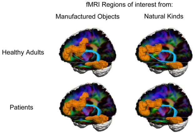
Figure 3.
White matter tractography in healthy adults and patients. Regions of interest (orange) were formed in left temporal-occipital cortex and prefrontal cortex based on fMRI results of healthy adults (Figure 1) showing common activations during judgments of manufactured objects or natural kinds. Streamline tractography between these regions is shown in light blue. RGB diffusion tensor imaging background shows water diffusion in tracts coursing in left-right (red), anterior-posterior (green), and superior-inferior (blue) orientations.

