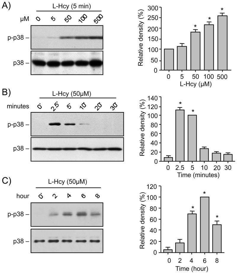
FIGURE 1.
Homocysteine induces biphasic pattern of p38 MAPK phosphorylation. Neuron cultures (A) were treated with different concentrations of L-homocysteine (L-Hcy) for 5 min, (B, C) were treated with 50 μM L-Hcy for the specified times. Equal amount of protein from each sample was processed for immunoblot analysis using anti-phospho-p38 (p-p38, upper panel) and p38 (lower panel) antibodies. Quantification of phosphorylated p38 MAPK by computer-assisted densitometry and Image J analysis is shown beside each immunoblot. Values are mean ± s.e.m. (n=3). * Significant difference from 0 min (p < 0.01).

