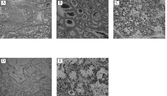
Figure 3.

A) The tumor consisted of papillary and cribriform pattern in the dilated mammary ducts (Haematoxylin and Eosin 50×); B) the tumor nests show microcystic structure with round spaces containing an eosinophilic secretion. Histological features mimic those of pregnant or lactational changes (Haematoxylin and Eosin 100×); C) immunohistochemical staining for periodic acid-Shiff (immunohistochemical analysis of periodic acid-Shiff, 400×); D) immunohistochemical staining for HHF35 (immunohistochemical analysis of HHF35, 100×); E) most tumor cells have vacuolated cytoplasm, and eosinophilic materials can be seen in the vacuoles (Haematoxylin and Eosin 400×).
