
Fig. 1.
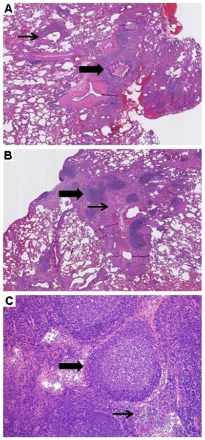
GLILD: Histologic findings. a Follicular bronchiolitis (thick arrow), with lymphocytic aggregates around an airway (thin arrow). (Lung, H&E, 100×, patient 2). b Focal lymphoid aggregates (thick arrow) within collagenized fibrous tissue (thin arrow). (Lung, H&E, 100×, patient 5). c Mediastinal biopsy reveals follicular hyperplasia with prominent germinal centers (thick arrow) and associated para-cortical hyperplasia. Interfollicular areas (thin arrow) are expanded. (Lymph node, H&E, 40×, patient 4)