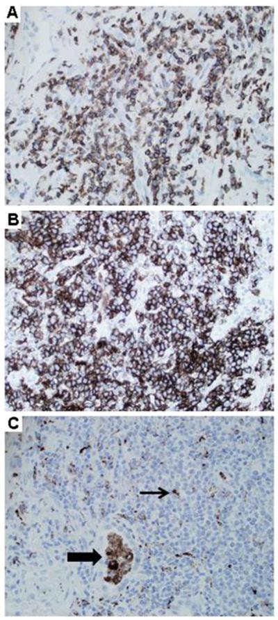
Fig. 2.

GLILD: Immunohistochemistry. a CD3 staining reveals frequent T cells. (Lung, 200×, patient 5). b CD20 staining demonstrates numerous B cells. (Lung, 200×, patient 5). c CD68 staining shows alveolar macrophages (thick arrow), and scattered dendritic cells (thin arrow) within lymphoid aggregates. (Lung, 200×, patient 5)
