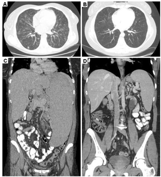
Fig. 4.
HRCT scans of the chest, abdomen in a patient with CVID, GLILD and a mutation in TACI. a Pre-treatment HRCT scan of the chest demonstrates diffuse pulmonary ground glass and nodular opacities. (Patient 6). b Improvement in pulmonary parenchymal abnormalities post-chemotherapy (Patient 6). c Pre-treatment splenomegaly extends to the iliac crest. (Patient 6; note: maximal spleen length shown). d Marked decrease in splenomegaly post-chemotherapy. (Patient 6; note: maximal spleen length shown)

