
Figure 1. Conservation of surface residues of the HIV-1 MA protein.
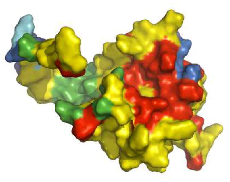
Color scheme: red = >99% conservative residue; yellow = >80%–99%; green = >50%–80%; cyan = >20%–50%; blue = 0%–20%. Residues surrounding a structural recess that serves as a binding site for PI(4,5)P2 are highly conserved as indicated by the red coloring.