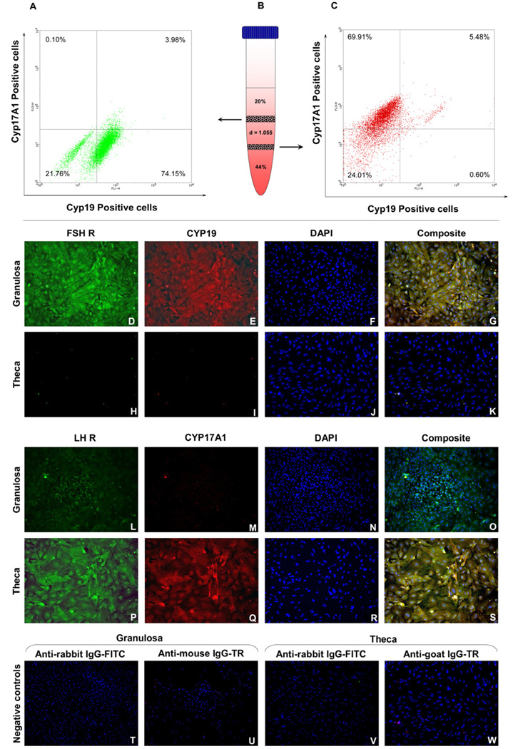
Fig 2.
Flow cytometric analysis of purity of isolated granulosa cells (A) retrieved from the first interphase of the discontinuous Percoll gradient (B) and theca cells (C) from the second interphase. Immuno-fluorescent staining for FSHR and CYP19 in granulosa (D–G) and theca cells (H–K); LHR and CYP17A1 in granulosa (L–O) and theca cells (P–S); Negative controls with secondary antibodies only on each cell type (T–W). Images acquired at 100 × magnification. FSHR – follicle-stimulating hormone receptor, LHR – luteinizing hormone receptor, CYP19 – aromatase, CYP17A1 – 17, 20 lyase, IgG – immunoglobulin G, FITC – Fluorescein isothiocyanate, TR – Texas Red.

