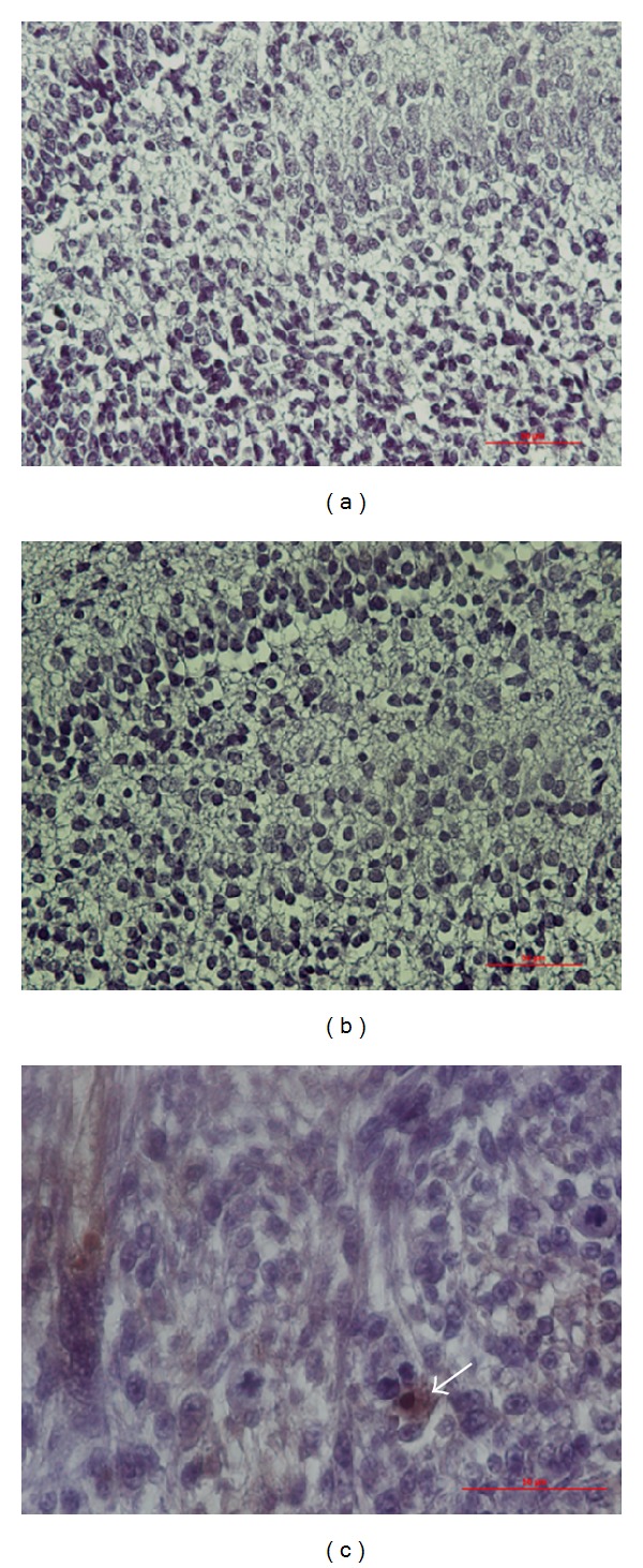
Figure 6.

OX-6 IHC staining in the DG. No OX-6 positive cells were identified in P1 D1D2 pups treated with either NS (a) or Dex (b). Brain tumor implant staining as a positive control showed OX-6 positive cells stained brown (c), arrow). Magnification, 400x; scale bar, 50 μm.
