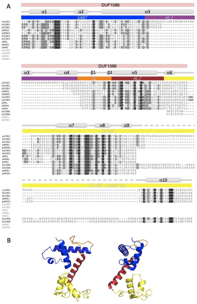
Figure 2.
PKL possesses a predicted DNA-binding domain that is similar to the DNA-binding domain of S. cerevisiae CHD1. (A) Alignment of scCHD1 with corresponding regions of CHD proteins from H. sapiens (hs), D. rerio (dr), A. thaliana (at), and the model grass species B. distachyon (bd). Specific regions of scCHD1 are marked as described in Ryan et al. (2011): SANT (blue), SLIDE (yellow), helical linker-1 (purple; HL1) and helical linker-2 (red; HL2) and β-linker (orange; βL). The region of CHD3 proteins that corresponds to DUF1086 is also marked. The extent of conservation of conserved residues is indicated by shading, and numbers in parentheses for CHD3 proteins indicate numbers of residues found in these remodelers that are not included in the region of conservation. (B) A cartoon representation of the predicted crystal structure of the putative DNA-binding domain of PKL is shown in two orientations. The domains are colored to reflect analogous domains indicated in scCHD1 in (A). Accession numbers for proteins used are provided in Table S1.

