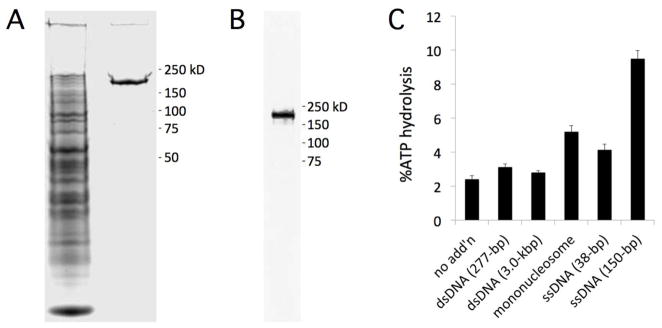
Figure 4.

Recombinant PKL-FLAG is a nucleosome-stimulated ATPase. (A) Extract from infected S2 cells was treated with anti-FLAG M2 affinity resin and applied to SDS-PAGE and stained with Coomassie Blue. The left lane is the unbound fraction and the right lane is purified PKL-FLAG. (B) Purified PKL-FLAG was analyzed by western blotting using an a-FLAG antibody. (C) Recombinant PKL-FLAG was incubated with ATP in the presence or absence of dsDNA, mononucleosomes, or ssDNA (indicated on x-axis). Hydrolysis of ATP was measured using a colorimetric assay and buffer alone was used as a reference for each sample. MW standards for panels A and B are indicated to right. Error bars in panel C represent SE calculated from 3 replicates.
