Fig. 3.
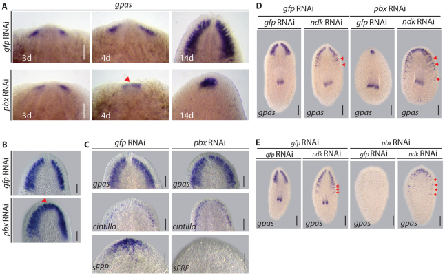
pbx is required for CG patterning. (A) By 3 dpa, CG primordia expressed the neural marker GPAS in the anterior blastema of regenerating trunk pieces in control (10/10) and pbx(RNAi) (10/10). At 4 dpa, these structures remained separate in control regenerates (8/8), whereas they started to coalesce in pbx(RNAi) (8/8) (red arrowhead). By 14 dpa, the CG was completely regenerated in controls (10/10), whereas in pbx(RNAi) regenerates the fused CG did not enlarge significantly (10/10). (B) Following longitudinal amputation the structure of the CG was correctly regenerated by 14 dpa in control lateral regenerates (12/12). Following pbx(RNAi), CG tissue was regenerated to the same extent as in controls; however, the structure was fused at the midline (17/17 fused, red arrowhead). (C) The CG structure was comparable in intact control (12/12) and pbx(RNAi) (14/14) planarians, as was the presence of cintillo-expressing sensory cells (8/8 and 8/8, respectively). In control planarians, sFRP was expressed at the anterior margin (13/13), whereas it was lost following pbx(RNAi) within 3 weeks of homeostasis (9/13). (D,E) In control gfp(RNAi) background, ndk(RNAi) resulted in expansion of the regenerated CG in 14 dpa tail and trunk pieces (17/17 control animals normal, 18/18 tails and 20/20 trunks in ndk(RNAi) expanded, red arrows). CG regeneration was not observed following pbx/gfp(RNAi) in tails (22/22) and was greatly reduced in trunks (20/20). Tail and trunk pieces regenerated mis-patterned CG at the anterior following combined pbx/ndk(RNAi) (20/20 animals). Scale bars: 100 μm.

