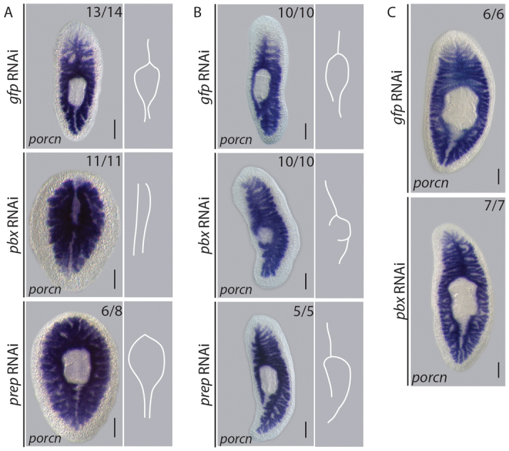
Fig. 5.

pbx coordinates AP patterning of the regenerating gut.
(A) The anterior gut branch was regenerated and the existing posterior gut branches re-patterned by 14 dpa in control tail pieces (14/14), revealed by gut-specific Smed-porcupine expression. The anterior gut branch failed to regenerate and the existing posterior gut did not re-model following pbx(RNAi) (11/11). The posterior gut remodeled in prep(RNAi) tail pieces; however, the anterior gut branch did not (8/8). Schematic representations of gut morphology summarize the phenotypes observed. (B) By 14 dpa, the posterior gut branch was restored in control lateral regenerates (10/10). The gut extended into the lateral blastema following pbx(RNAi); however, patterning of the posterior branch was not regenerated (10/10). Regeneration of the posterior gut was comparable with controls following prep(RNAi) (10/10). Schematic representations of gut morphology summarize the phenotypes observed. (C) Gut morphology is maintained during 3 weeks of homeostasis following pbx(RNAi) (17/17), resembling controls (16/16). Scale bars: 200 μm.
