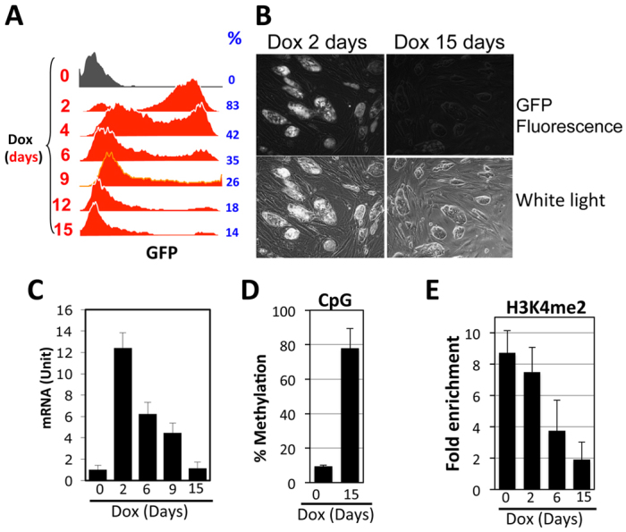
Fig. 3.

Effects of rtTA on the CMV-GFP transgene in ES cells. Cells were cultured in the presence of Dox (1 μg/ml) for various days before analysis. (A) FACS analysis of GFP expression. Dox stimulation times and percentage of GFP-expressing cells are shown at left and right, respectively. (B) Micrographs of ES cells following 2 (left) and 15 (right) days of Dox exposure. (C) GFP mRNA levels, normalized to β-actin mRNA. (D,E) CpG methylation and H3K4me2 at the CMV promoter, as described in Fig. 2. The values were averaged from two and three independent experiments for D and E, respectively. Error bars indicate variations between the independent trials.
