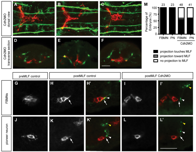
Fig. 8.
Depletion of Cdh2 reduces interactions between the FBMNs and MLF. Maximum projections of Tg(zCREST1:membRFP) embryos stained using zn-12. (A-C) Dorsal views of Cdh2MO-injected embryos at (A) 20 hpf, (B) 22 hpf and (C) 24 hpf show reduced interaction between FBMNs (red) and the MLF (green). (D-F) Transverse sections of Cdh2-depleted embryos at r5 show decreased interactions between the MLF and FBMNs at (D) 20 hpf, (E) 22 hpf and (F) 24 hpf. (G-L′) High-magnification images of transverse sections at 19.5 hpf. Yellow arrowheads indicate the LLF. (G) Before the MLF is present, FBMNs send projections in random directions. (H,H′) Once the MLF (white arrowhead) is present, FBMNs send a projection to the MLF (arrow). (I,I′) In Cdh2-depleted embryos, FBMNs in the midline do not send projections to the MLF. (J) The pioneer neuron, before the MLF has entered r5, projects randomly. (K,K′) Once the MLF is present, the pioneer neuron sends a projection towards it. (L,L′) In Cdh2-depleted embryos, the pioneer has reduced interactions with the MLF. (M) Percentage of embryos with neural projections contacting the MLF. Scale bar: 20 μm.

