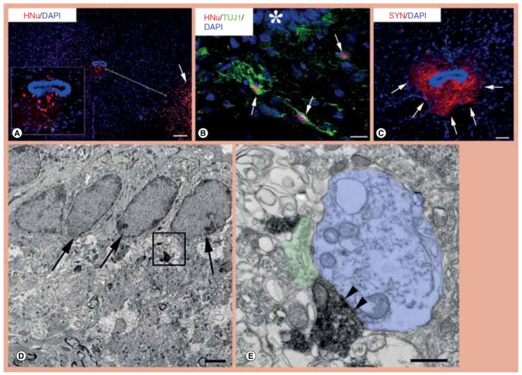
Figure 1. Migration of transplanted human neural stem cells from the inoculation site to the central canal zone and differentiation into neurons with mature synapses.

(A) Transplanted neural stem cells (NSCs) were attracted to the central canal by crossing the distance between the inoculation site in the ventral horn (red, lower right, arrow) and central canal. Inset is a magnification of the central canal zone with migrated NSC-derived cells. (B) NSCs that have migrated from the initial transplantation site to the ventral central canal (asterisk) differentiate extensively into TUJ1-positive neurons (green, arrows). (C) In this section that is adjacent to the section photographed for (A), NSCs that had migrated in the central canal zone are shown to generate dense human-specific SYN-positive terminals (arrows). (D & E) These electron photomicrographs taken from a human SYN-labeled section show SYN-positive terminals apparently derived from differentiated human NSCs next to ependymal cells (D, arrows). (E) Enlargement of the framed area in (D) to show the ultrastructure of a human NSC-derived terminal replete with synaptic vesicles, mitochondria and terminal membrane specializations (arrowheads) forming a synapse with an unlabelled, possibly dendritic, postsynaptic structure (blue); the human-derived synaptic terminal is juxtaposed to an unlabeled host terminal (green) on the same postsynaptic structure. Scale bars: (A) 100 μm; (B) 20 μm; (C) 25 μm; (D) 150 nm; (E) 50 nm.
DAPI: 4’,6-diamidino-2-phenylindole; HNu: Human nuclear protein antibody; SYN: Synaptophysin; TUJ1: Neuronal class III β-tubulin.
