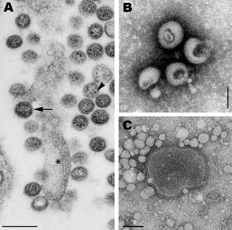
Figure 1.
A) Transmission electron micrograph of an ultrathin section of Vero cells infected with Cygnet River virus (CyRV) from a Muscovy duck, Australia. Arrow, virus budding from the plasma membrane; arrowhead, sand-like structures. *Host cell projection. Scale bar = 200 nm. B, C) Transmission electron micrographs of CyRV prepared by negative-contrast electron microscopy. Scale bars = 100 nm. Preparations were derived from supernatant of CyRV-infected Vero cells (B) and from allantoic fluid of CyRV-infected eggs (C).

