Figure.
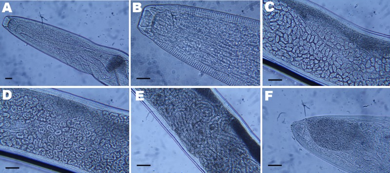
Light micrographs of Thelazia callipaeda showing A) posterior and B) anterior portion with cephalic end and buccal capsule; C) anterior portion containing embryonated eggs; D) middle portion containing rounded first-stage larvae; E) posterior portion containing first-stage larvae; F) caudal end. Scale bars = 25 µm.

