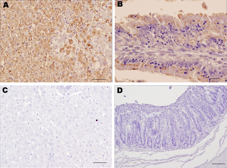
Figure 2.
Results of immunohistochemical staining using monoclonal antibody 6G2 and the ABC complex technique of liver and intestine samples from young rabbits infected with rabbit hemorrhagic disease virus (RHDV) isolate RHDV-N11 and control rabbits. A) Liver of RHDV-N11–infected rabbit. Hepatocytes show intense 6G2-specific immunolabeling. Scale bar = 50 µm. B) Intestinal villi in small intestine of RHDV-N11–infected rabbit. Areas of focal necrosis and epithelial cells show strong immunolabeling. Scale bar = 20 µm. C) Liver of control rabbit. Hepatocytes do not show positive immunolabeling. Scale bar = 50 µm. D) Epithelial cells of intestinal villi of control rabbit do not show positive results on staining. Scale bar = 100 µm.

