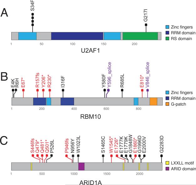
Figure 3. Somatic mutations of lung adenocarcinoma candidate genes U2AF1, RBM10, and ARID1A.
(A) Schematic representation of identified somatic mutations in U2AF1 shown in the context of the known domain structure of the protein. Numbers refer to amino acid residues. Each rectangle corresponds to an independent, mutated tumor sample. Silent mutations are not shown. Missense mutations are shown in black. (B) Schematic of somatic RBM10 mutations. Splice site mutations are shown in purple; truncating mutations are shown in red. Other notations as in (A). (C) Schematic of somatic ARID1A mutations. Notations as in (A) and (B).

