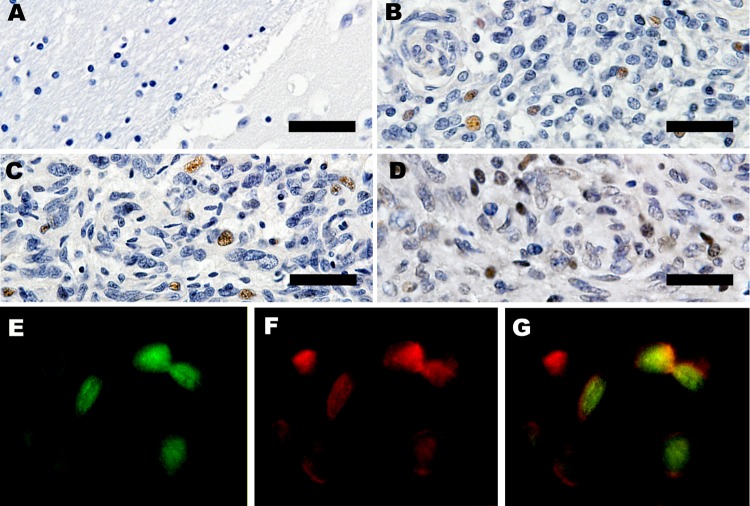
Figure 6.
Expression of large T-antigen (LT-Ag) and p53 within a subset of tumors. A) Control, immunohistochemical analysis of frontal lobe of normal raccoon brain tissue. Astrocytes in this image and throughout the section were not immunoreactive for LT-Ag. Original magnification ×40. B, C) Immunohistochemical analysis for raccoon no. 2 (Rac2) and Rac3. LT-Ag is expressed within the nuclei of a subset of neoplastic astrocytes in 2 independent tumors. Original magnification ×40. D) Immunohistochemical analysis for Rac3. p53 is present within the nuclei of a subset of tumor cells. Original magnification ×40. Scale bars for panels A–D = 100 μm. E–G) Immunofluorescence of Rac4. p53 co-localizes with LT-Ag within nuclei of neoplastic cells. Original magnification ×60.

