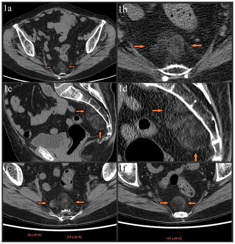
Figure 1.
79 year old female with presacral myelolipoma. The CT was performed on a GE® 64-slice CT scanner. All images are from the patient’s initial noncontrast bone algorithm pelvic CT with standard soft tissue windows (width = 400, center = 50). 2mm slices were acquired, using 120 kVp and variable mAs, which ranged from 614 mAs to 633 mAs for the provided axial images.
Figures 1a–1f: Axial (figure 1a, magnified in figure 1b) and sagittal (figure 1c, magnified in figure 1d) images show incidental 5.8 × 2.9 × 4.8 cm lobulated, heterogeneous presacral mass without evidence of erosion/invasion of the anterior sacrum (orange arrows). The mass spans from S3 to S5 and is composed of fat and soft tissue elements. Precise measurement of the soft tissue elements’ attenuation is difficult due to significant admixture, but attempts show a range from about −10 HU to about 20 HU, with a single area of higher (mid 30 HUs) attenuation in the lesion’s right lateral margin (image 1e). The fat elements have attenuation values of about −100 HU (image 1f).

