Figure 2.
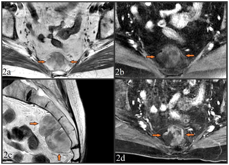
MR images from a 79 year old female with presacral myelolipoma. All images were acquired on a GE® 1.5 Tesla MR scanner.
Figure 2a. Pre-contrast T1-weighted fast spin echo (FSE) axial image (TR=500.0, TE=9.0, TI=0.0, FA=90.0) at the level of the midsacrum demonstrated a 6.4 × 3.1 × 5.7 cm lobulated presacral mass with mixed fat/soft tissue signal (arrows). No bony invasion was seen.
Figure 2b. Pre-contrast fat-suppressed T1-weighted FSE axial image (TR=516.7, TE=7.7, TI=0.0, FA=90.0) taken at the same level, showed loss of signal intensity in the areas which were previously iso-intense to fat, providing further confirmation of a significant fat component of the mass.
Figure 2c. Pre-contrast T2-weighted FSE image (TR=5800.0, TE=119.8, TI=0.0, FA=90.0) in the sagittal plane to provide craniocaudad visualization of the 6.4 × 3.1 × 5.7 cm mass which spanned from S3 to S5. The mass was again noted to be lobulated, of mixed fat/soft tissue constituency, and without bony invasion.
Figure 2d. Post-contrast (15cc of gadopentetate dimeglumine) T1-weighted fat-suppressed liver acquisition with volume acquisition (LAVA) sequence axial image (TR=4.4, TE=2.1, TI=7.0, FA=12.0) taken at the same level of images 2b/2c showed enhancement of the non-fatty soft tissue elements, and redemonstrated loss of signal in the mass’ fatty elements.

