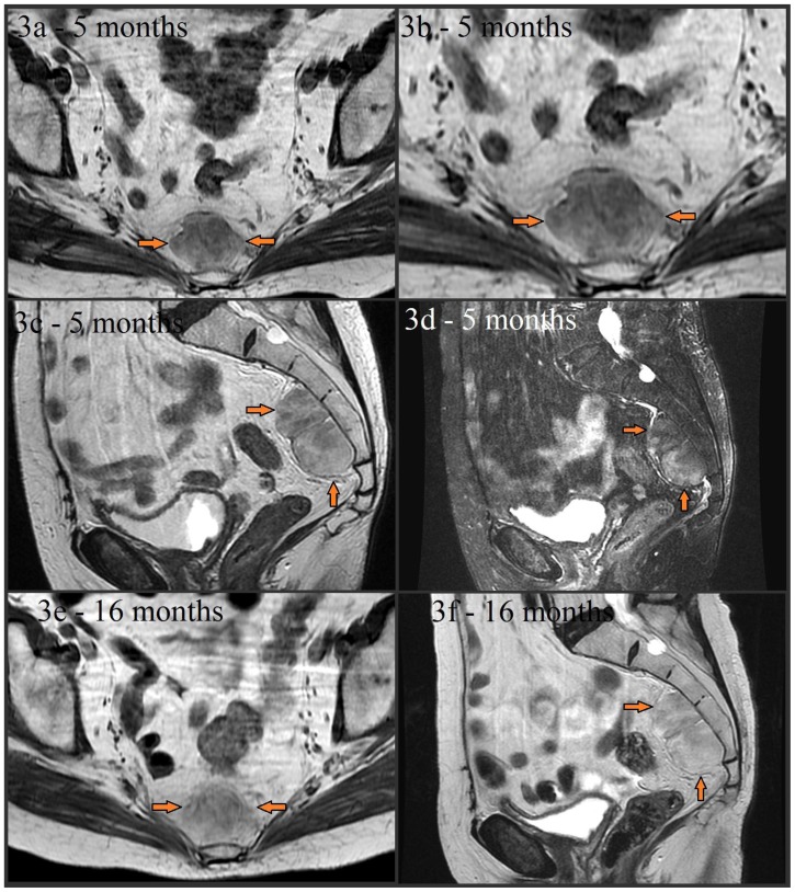
Figure 3.
Noncontrast MR images from a 79 year old (80 year old at the time of images 3e and 3f) female with presacral myelolipoma. All images were acquired on a GE® 1.5 Tesla MR scanner.
Follow-up axial noncontrast T1-weighted FSE (image 3a and magnified in 3b, TR=400.0, TE=8.5, TI=0.0, FA=90.0), noncontrast sagittal T2-weighted (image 3c, TR=3166.7, TE=122.1, TI=0.0, FA=90.0), and noncontrast sagittal T2-weighted fat-suppressed (image 3d, TR=3450.0, TE=122.1, TI=0.0, FA=90.0) images taken about 5 months later showed stability of the presacral mass (arrows) without significant interval change in size, appearance, or signal characteristics. The mass is again noted to be lobulated, well-circumscribed, and have mixed soft tissue and fat composition (with signal dropout on fat-suppressed image 3d). A lack of sacral invasion is again seen. Noncontrast T1-weighted FSE (image 3e, TR=433.3, TE=7.5, TI=0.0, FA=90.0) and noncontrast sagittal T2-weighted (image 3f, TR=5450.0, TE=119.9, TI=0.0, FA=90.0) images obtained 16 months later also showed continued overall stability.

