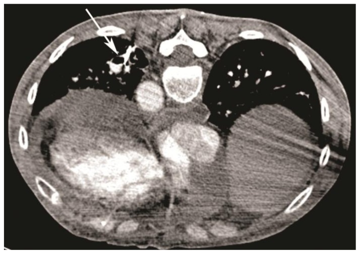
Figure 3.
59-year-old woman with scleroderma and a solitary lung nodule, which turned out to be an aneurysm after biopsy. Repeat CT angiogram (64-Channel CT scanner. Sensation 64, Siemens Medical Solutions, 120 kV, 306 mAs, 3 mm reformation, 80 c.c. of 300 mg/ml iodine concentration non-ionic contrast) after injection of 2 cc of air to better delineate the walls (white arrow, patient is on prone position) demonstrating contrast filling and pooling into the cavity suggestive for a patent aneurysm.

