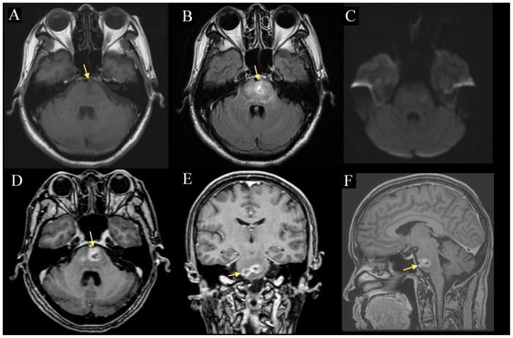
Figure 1.
57-year-old man with radiation necrosis of the pons. Contrast-enhanced MP-RAGE (1D-F) MRI demonstrates two well-circumscribed peripherally contrast-enhancing lesions (arrow) in the pons measuring 14 and 15 mm respectively. The lesions are hypointense on pre-contrast T1 (1A) and are associated with diffuse FLAIR (1B) edema that extends into the medulla and right inferior cerebellar peduncle. There is no obvious mass effect and no extension of the lesion beyond the boundaries of the pons, notably into the cerebellopontine angle or the prepontine cistern. DWI (1C) showed no diffusion abnormality. (A: 1.5 Tesla, TR 400ms, TE 16ms, slice thickness 5.0mm, B: TR 8602ms, TE 129.3ms, slice thickness 5.0mm, C: TR 10000ms, TE 98.3, slice thickness 5.0mm, D: TR 8.4ms, TE 2.6ms, slice thickness 1.6mm. E-F: TR 8.4ms, TE 2.6ms, slice thickness 1.5mm; A-C without contrast, D-F with 10mL of gadopentate dimeglumine (Magnevist))

