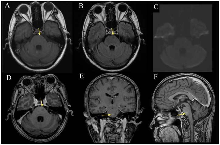
Figure 3.
57-year-old man with radiation necrosis of the pons. Compared to the prior study (figure 1), there is interval size reduction and normalization (arrow) in the T1 (3A) hypointensity, FLAIR (3B) pontine signal abnormality and the associated contrast enhancement on MP-RAGE (3D-F). DWI (3C) continues to show no diffusion abnormality. There is no obvious mass effect of the pontine and cerebellar peduncle lesions and no extension beyond the boundary of the brainstem. (A: 1.5 Tesla, TR 400ms, TE 16ms, slice thickness 5.0mm, B: TR 8602ms, TE 129.4ms, slice thickness 5.0mm, C: TR 10000ms, TE 98.3ms, slice thickness 5.0mm, D: TR 8.4ms, TE 2.6ms, slice thickness 1.6mm. E-F: TR 8.4ms, TE 2.6ms, slice thickness 1.5mm; A-C without contrast, D-F with 13mL of gadopentate dimeglumine (Magnevist))

