
Figure 3.
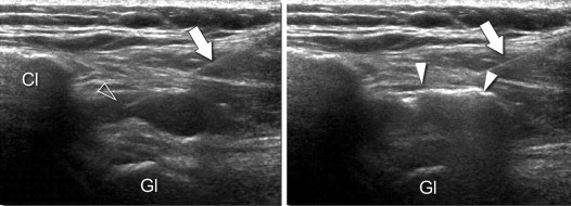
Superior approach. Oblique coronal US scans (see Fig. 1, the central image). Left: image showing the needle correctly positioned before injection. Arrow = needle; arrowhead = the needle tip inside the subacromial-subdeltoid bursa which is distended by synovial fluid. Right: US scan taken during corticosteroid injection (white arrowheads); note that the drug is being correctly injected into the bursa. Cl = clavicle; GL = glenoid.