
Figure 4.
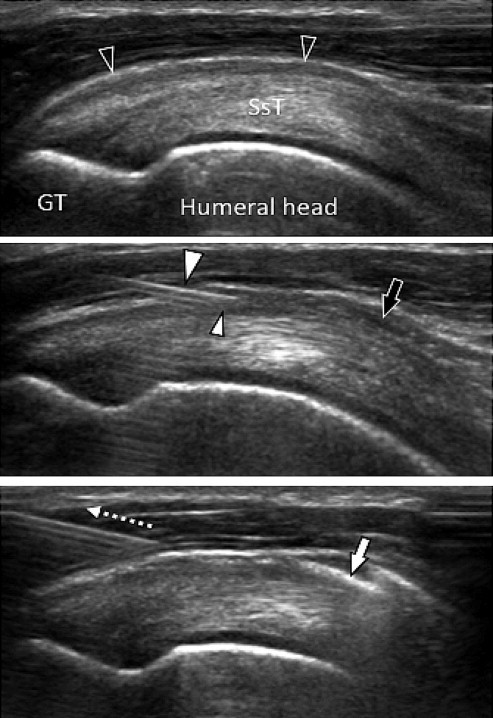
Lateral approach. Oblique coronal US scans (see fig. 1, the right image). Above: image taken before corticosteroid injection; note the significant thickening of the subacromial-subdeltoid bursa (empty arrowheads). SsT = supraspinatus tendon; GT = greater tuberosity of the humerus. Center: The needle (large white arrowhead) is correctly positioned; the tip (small white arrowhead) is inside the subacromial-subdeltoid bursa. A small amount of injected drug (black arrow) is visible inside the subacromial-subdeltoid bursa. Bottom: When the corticosteroid injection is completed the needle is removed (dotted arrow); air microbubbles (white arrow).